Research
Localization of centromeric histone CENP-A during cell division in P. patens.
This figure is adapted from the following publication:
Kozgunova E., Nishina M., Goshima G. Kinetochore protein depletion underlies cytokinesis failure and somatic polyploidization in the moss Physcomitrella patens. eLife 8:e43652 (2019). https://doi.org/10.7554/eLife.43652
Microfluidic device for high resolution cytoskeleton imaging
Microfluidic chip or device is a set of micro-channels molded into a material (PDMS) in which plant cells can be grown without any adverse effects. We can precisely modulate the channel environment by controlling liquid flow in the channels, introducing or wash-out chemical compounds, and observing the cellular response. Furthermore, microdevice offers excellent properties for high-resolution imaging, such as TIRF or VAEM imaging, and can be used to visualize protein dynamics at the molecular level.
We use microdevices for high-resolution microtubules (MTs) imaging in the protonema cells of moss P. patens and testing the effect of various chemical compounds on cytoskeleton dynamics.
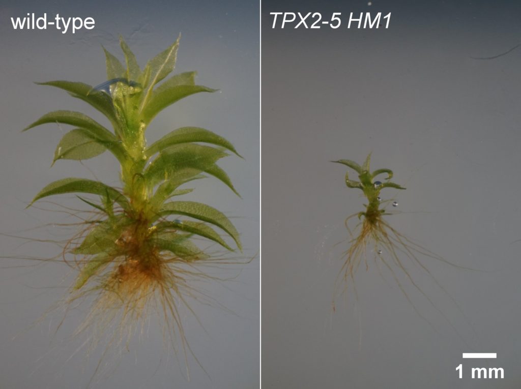
Spindle motility has dramatic consequences for gametophore (leafy shoot) development in P.patens
This figure is adapted from the following publication:
Kozgunova, E., Yoshida, M.W., Reski, R. et al. Spindle motility skews division site determination during asymmetric cell division in Physcomitrella. Nat Commun 13, 2488 (2022). https://doi.org/10.1038/s41467-022-30239-1
Plant kinetochore and polyploidy
The kinetochore is a macromolecular complex that directs chromosome segregation by connecting chromosomes and spindle microtubules. Previously, we conducted a comprehensive screen of all major kinetochore components in P. patens and made an unexpected discovery: defects in chromosome segregation, completely blocked cytokinesis in a spatio-temporal manner. The resulting cells had two nuclei and eventually developed into polyploid plants. In future, we plan to further investigate relationship between polyploidy and kinetochore in plants.
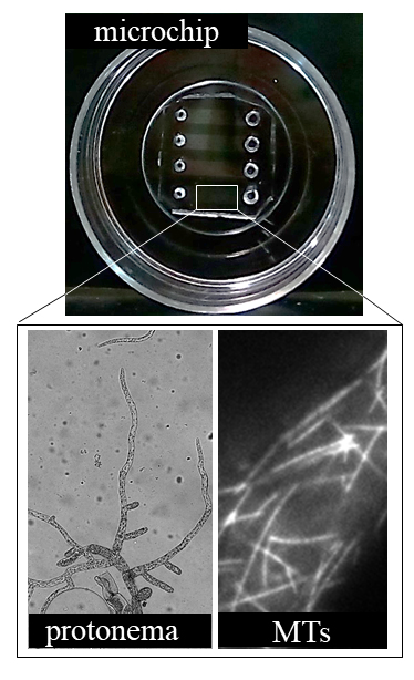
Microfluidic device for high-resolution microtubule imaging in protonema cells of moss P. patens.
This figure is adapted from the following publication:
Kozgunova, E., Goshima, G. A versatile microfluidic device for highly inclined thin illumination microscopy in the moss Physcomitrella patens. Sci Rep 9, 15182 (2019). https://doi.org/10.1038/s41598-019-51624-9
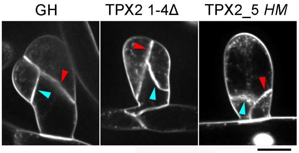
Spindle formation and cell division plane determination
In animals, mitotic spindle positioning determines the division site during asymmetric cell division. By contrast, in plants, a pre-mitotic structure known as the pre-prophase band (PPB) marks the site of future cell division. For many years, it was thought that the plant cell division site was determined before mitosis and no evidence of active spindle positioning in plant cells was discovered until now. In the recent work, we characterized the function of the microtubule-associated protein Targeting protein for Xklp2 (TPX2) during mitosis in P. patens and isolated a hypomorphic mutant of TPX2 that showed drastic spindle motility leading to a reversed cell ratio in asymmetric cell division and impaired organ development. This discovery laid the foundation for this project: understanding the role of spindle positioning in plant development from an evolutionary perspective.
Talk to us
Have any questions? We are always open to talk about new projects, collaboration opportunities and how we can help you.